16+ Plantar Foot Surface
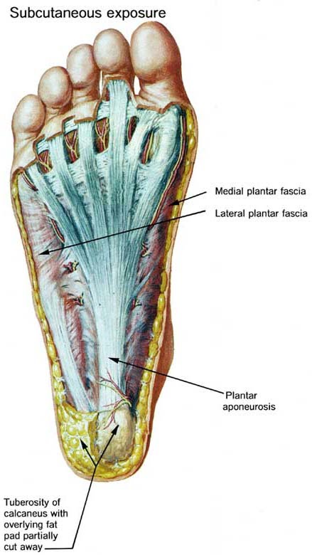
Web The plantar fascia PF is the most superficial structure in the sole of the foot originating from the plantar surface of the calcaneus at the medial calcaneal tubercle and attaching to the plantar surfaces of the five metatarsal heads and proximal phalanges of the toes. Web Lateral plantar muscles inferior view The lateral chamber formed by the plantar fascia contains three muscles.

Plantar Fasciitis And Other Abnormalities Of The Plantar Fascia Radiology Key
What are the 3 portions of the plantar aponeurosis.

. An overview of foot pain generally including more detailed discussions of foot anatomy and biomechanics and how to conduct a history and examination of the patient with foot. Introduction The foot is a complex structure comprised of over 26 bones 30 joints numerous tendons ligaments and muscles responsible for our ability to stand upright supporting the weight of the entire body and provides the base for the mechanism for bipedal gait. Plantar fasciitis PLAN-tur fas-e-I-tis is one of the most common causes of heel pain.
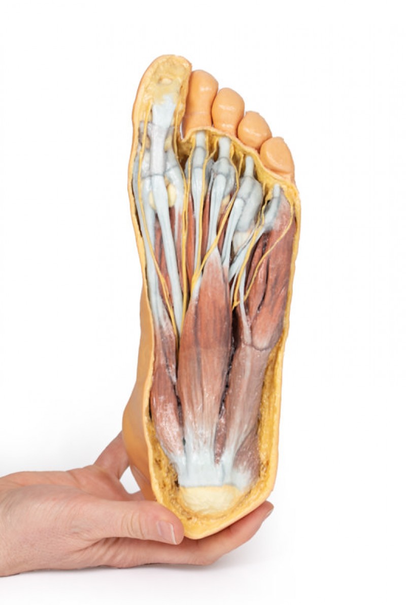
It is embedded in a tendon at the head of the bone the part closest to the big toe. Web - Plantar muscles of the foot are divided into 4 layers. Web This image shows the anatomy of the plantar foot and is labeled with corresponding identification tags.
Plantar fasciitis can cause intense heel pain. Flexor digitorum brevis FDB abductor digiti minimi. This tissue originates deep within the plantar surface of the calcaneus heel bone and covers the distance to the base of each of the five toes.
Web The plantar fascia is not a nerve tendon or muscle but rather a strong fibrous tissue Figure 16. It is covered with hairless usually nonpigmented skin that is especially thickened and provided with epidermal ridges over the weight-bearing areas. Web A comprehensive literature review is presented to better define available reconstructive options for resurfacing the plantar foot.
The compartment comprises numerous short foot muscles in different layers. Plantar Surface of Foot. The muscles lying within the medial group form a bulge referred to as the ball of the big toe.
Click the card to flip. The PubMed Embase and Scopus databases were queried for published articles. Web Tendon disorders along the plantar aspect of the foot may lead to significant symptoms but are often clinically misdiagnosed.
Web the inferior aspect or bottom of the foot much of which is in contact with the ground when standing. Click the card to flip. Web In metatarsalgia a directed diagnostic work-up to find the cause is mandatory including a search for excessive mechanical stress due to abnormal foot posture neuropathic pain rheumatoid arthritis aseptic bony necrosis or malignant disease.
Familiarity with the normal anatomy of the plantar tendons and its appearance at magnetic resonance MR imaging and ultrasonography US is essential for recognizing plantar tendon disorders. A systematic literature search was performed to identify articles relating to reconstruction of the plantar skin and soft tissue. Web Sesamoid bones.
Extrinsic Muscles of the Foot Peroneus Longus. Together they form the central surface of the foot sole. Web The plantar surface was divided into eight anatomical regions including great toe T1 lesser toes T2-5 first metatarsal M1 central forefoot M23 lateral forefoot M45 midfoot MF.
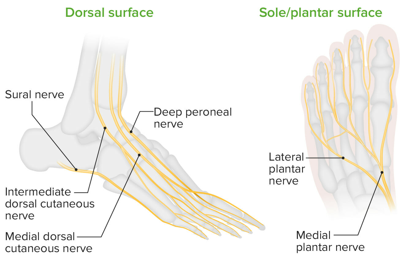
In this article we present the clinical findings of the diseases that most commonly cause plantar pain the diagnostic procedures available and an overview of the treatment options. Web Pain on the plantar surface of the foot subsumes constellations of symptoms arising from various underlying conditions. Web Identify the following structures passing into the plantar surface of the foot posterior to the malleolus in the tarsal tunnel from anterior to posterior.
It consists of a central medial and lateral part. These are two small oval-shaped bones beneath the first metatarsal on the underside plantar surface of the foot. It contributes to the surface anatomy of the medial sole of the foot and is easy to palpate.
Web Plantar cutaneous afferents transmit spatial and temporal feedback concerning the pressure variations and skin stretch exerted on the soles of the feet 58 63 179. It helps support the arch of the foot and has an important role. - first subfascial layer consists from lateral to medial of the ADM FDB.
Their muscle bellies form the surface of the lateral foot sole ball of the little toe. In response to this sensory feedback corrective postural reactions are evoked by postural muscles 148 176 177. Web The plantar fascia which surrounds all muscles of the sole of the foot consists of three chambers.
Web Go to. Lateral central and medial. Imaging studies and pedobarography are needed.
Planta pedis TA plantar region regio plantaris plantar surface of foot. Web Plantar fasciitis is one of the most common conditions causing heel pain. Tendon of tibialis posterior tendon of flexor digitorum longus posterior tibial artery posterior tibial veins tibial nerve tendon of flexor hallucis longus.
When the foot rolls off the ground during walking the toes dorsiflex and pull on the plantar fascia. - first two layers originate from calcaneal tuberosity and the other two from the metatarsal shafts. Web The intrinsic muscles of the foot are further divided into dorsal and plantar groups and there are four layers of the plantar muscles.
The plantar fascia attaches to the heel bone calcaneus and to the base of the toes. Web This topic reviews the common causes of forefoot pain in the adult including descriptions of important conditions and a discussion of how to reach a diagnosis. Web Plantar fasciitis is an inflammation of the fibrous tissue plantar fascia along the bottom of your foot that connects your heel bone to your toes.
They are all innervated by the lateral plantar nerve S1-S2 a branch of the tibial nerve. Web Anatomy and supply Central plantar muscles The central muscles of the foot sole lie within the central compartment between the muscles of the big and little toe. Most superficial of all the layers.
- plantar fascia is comparable to palmar aponeurosis. It involves inflammation of the plantar fascia a tough fibrous band of tissue that runs along the sole of the foot.

Foot Structures Of The Plantar Surface Lower Limb Anatomy Series 1 0 3d Anatomy Series Shop Erler Zimmer
Phantom Dorsal Night Splint Med Spec

Pain On The Plantar Surface Of The Foot Review Article Iaom Us
Hyperkeratosis Of The Foot Part 1 Document Gale Onefile Health And Medicine

Foot Structures Of The Plantar Surface Lower Limb Anatomy Series 1 0 3d Anatomy Series Shop Erler Zimmer
Pain On The Plantar Surface Of The Foot 08 02 2019

Plantar Surfaces Hi Res Stock Photography And Images Alamy
Foot Anatomy Concise Medical Knowledge

The Foot The Superficial Plantar Fascia And Plantar Fat Pad Youtube

The Axes And Quadrants Of The Plantar Surface Of The Feet 1 Quadrant Download Scientific Diagram

Plantar And Dorsal Surfaces Of The Foot Showing The Deep Plantar Download Scientific Diagram
Anatomy Of Plantar Foot Surface Levin And Chellen Chiropractic

Foot Structures Of The Plantar Surface Lower Limb Anatomy Series 1 0 3d Anatomy Series Shop Erler Zimmer

Foot Structures Of The Plantar Surface Lower Limb Anatomy Series 1 0 3d Anatomy Series Shop Erler Zimmer
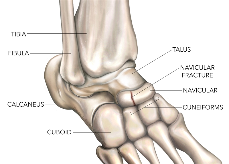
Sports Injury Bulletin Anatomy Navicular Stress Fracture A High Impact Risk For Young Athletes
Foot Plantar Surface Superficial Dissection On The Dorsum
The Foot The Superficial Plantar Fascia And Plantar Fat Pad Youtube